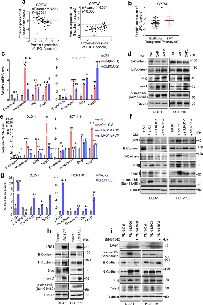
Fig. 3. CAFs-induced LRG1 promotes EMT.
a Correlation analysis between E-cadherin/N-cadherin protein expression and LRG1 expression in CRC primary tumors in CPTAC database. b Graph showing LRG1 protein expression in CRC primary tumor with two different integrated phenotypes (epithelial versus EMT) based on data from CPTAC. c EMT associated markers were analyzed by qRT-PCR in DLD-1 and HCT-116 upon treatment with control medium or CM from two individual CAF. d EMT associated markers and phosphorylated Smad1/5(ser463/465) were analyzed by western blot in DLD-1 and HCT-116 upon treatment with control medium or CM from two individual CAF. e DLD-1 and HCT-116 with or without silencing of LRG1 were treated with control medium or CM from CAF. Expression of EMT-associated markers was measured by qRT-PCR. f DLD-1 and HCT-116 with or without silencing of LRG1 were treated with control medium or CM from CAF. Expression of EMT-associated markers and phosphorylated Smad1/5(ser463/465) was measured by western blot. g Expression of EMT-associated markers was measured by qRT-PCR in DLD-1 and HCT-116 with or without ectopic expression of LRG1. h Expression of EMT-associated markers and phosphorylated Smad1/5(ser463/465) was measured by western blot in DLD-1 and HCT-116 with or without ectopic expression of LRG1. i DLD1 and HCT-116 in the presence or absence of LRG-1 overexpression were pre-treated with SB431542, a TGFβ-Smad pathway inhibitor, or not. Expression of LRG1, phosphorylated Smad1/5 (ser463/465) and EMT-associated markers were measured by western blot. Error bars represent SD; n = 3. *P < 0.05, **P < 0.01, ***P < 0.001, ****P < 0.0001.

