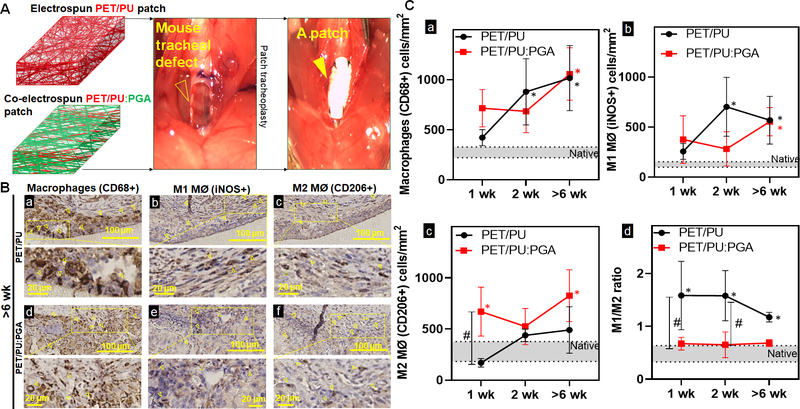
Figure 2. Macrophage quantification and phenotype in synthetic tracheal scaffolds.
A. Repair of an anterior tracheal defect (empty arrow head) with a synthetic patch (arrow head) made of either electrospun PET/PU or co-electrospun PET/PU:PGA B. Immunohistochemistry against macrophages (CD68+) (a, d), M1 macrophages (MØ) (iNOS+) (b, e), and M2 MØ (CD206+) (c, f) cells to indicate macrophage infiltrates in the submucosa over PET/PU and PET/PU:PGA patches were performed on serial axial sections. Representative images of implants >6weeks post-surgery shown. (1wk and 2wk sections not shown). Positive staining of macrophages is highlighted by the arrowheads. Scale bar=100 μm. C. Data was represented as macrophages (CD68+) (a), M1 MØ (iNOS+) (b), M2 MØ (CD206+) (c) macrophages per mm2 of the graft and the ratio of macrophage phenotype M1 (iNOS+)/ M2 (CD206+) (d) in the patch. Macrophage counts in native phenotypes are indicated by the grey bars. (n=4; ANOVA with Tukey’s test; * p<0.05 compared to native; # p<0.05 between PET/PU and PET/PU:PGA; Error bars represents the standard deviation)

