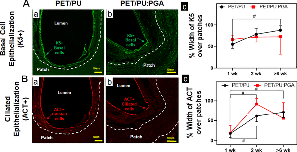
Figure 3. Epithelialization of synthetic PET/PU and PET/PU:PGA grafts.
A. Immunofluorescent staining for K5 to identify basal epithelial progenitor cells over synthetic patches were performed. Representative immunofluorescent images of K5+ basal cells over implanted PET/PU (a) and PET/PU:PGA (b) are shown. (c) Quantification of K5+ basal cell-coverage luminal to the patches were performed. (n=5; Kruskal-Wallis test; # p<0.05; Error bars represent standard deviation). B. Immunofluorescent staining against ACT antibodies to identify ciliated epithelial cells over synthetic patches were performed. Representative immunofluorescent images of ACT+ epithelial cells over implanted PET/PU (a) and PET/PU:PGA (b) are shown. (c) Quantification of ciliated epithelial cells (ACT+) luminal to the patches were performed. (n=5; ANOVA with Tukey’s test;; # p<0.05; Error bars represents the standard deviation)

