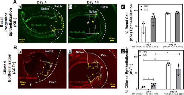
Figure 5. TETG epithelization is enhanced by macrophage depletion at the early time point.
A. Immunofluorescent staining against K5 antibodies to identify basal epithelial progenitor cells over PET/PU patches in PBS (a) and CCL (b) treated mice. The working area is delineated by the white # sign between the white arrows. The region of K5 coverage is denoted by yellow * between the yellow arrows. (c) Quantification of K5+ basal cell epithelization of the graft. (n=3, Kruskal-Wallis test; Error bars represents the standard deviation) B. Immunofluorescent staining against ACT antibodies to identify ciliated epithelial cells over PET/PU patches in PBS (a) and CCL (b) treated mice. The working area is delineated by the white # sign between the white arrows. The region of ciliated epithelialization is denoted by yellow * between the yellow arrows. (c) Quantification of ciliated epithelization (ACT+) luminal to the patches. (n=3; ANOVA with Tukey’s test; * p<0.05; # p<0.05; Error bars represents the standard deviation)

