FIG. 4.
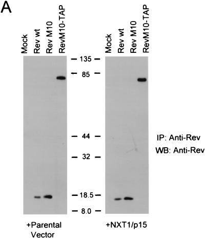
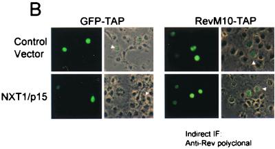
Expression of NXT1 does not alter the steady-state localization of M10-TAP or dramatically affect RevM10-TAP expression levels. (A) IP-Western blot (WB) analysis of proteins from transfected cells. Rev and RevM10-TAP fusion proteins were immunoprecipitated from lysates of transfected cells using an anti-Rev polyclonal antibody and separated using SDS-PAGE. Western blot analysis was performed with an anti-Rev monoclonal antibody (3H6) (45). Blots were visualized with ECL. Positions of commercial molecular weight standards (103 Bio-Rad) are indicated. (B) Fluorescence microscope analysis of GFP-TAP and RevM10-TAP proteins. Transfected CMT-3/COS cells were fixed and permeabilized. RevM10-TAP-expressing cells were stained using a primary anti-Rev rabbit polyclonal antibody and an Alexafluor-488-conjugated secondary antibody (Molecular Probes). Cells were visualized by fluorescence microscopy, and representative fields were photographed. wt, wild type. IF, immunofluorescence.


