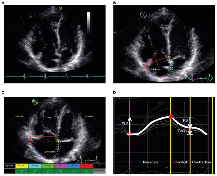
Figure 1.
Illustration of the assessment of RA strain. (A) First, the RV-focused apical four-chamber view was used with selection of the cardiac cycle and adjustment of the electrocardiogram (to R-wave). (B) Second, the RA endocardial border was traced as the region of interest, covering the RA lateral wall, roof, and septal wall. (C) Third, processing provided an overview wherever speckle tracking was feasible for the selected regions. (D) Fourth, the different phases were identified and the strain values determined. PACS, peak active contraction strain; PLS, peak longitudinal strain; PS, passive strain; RA, right atrial; RV, right ventricular.

