FIG. 7.
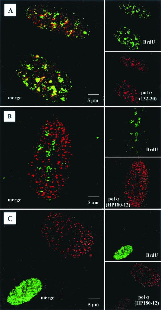
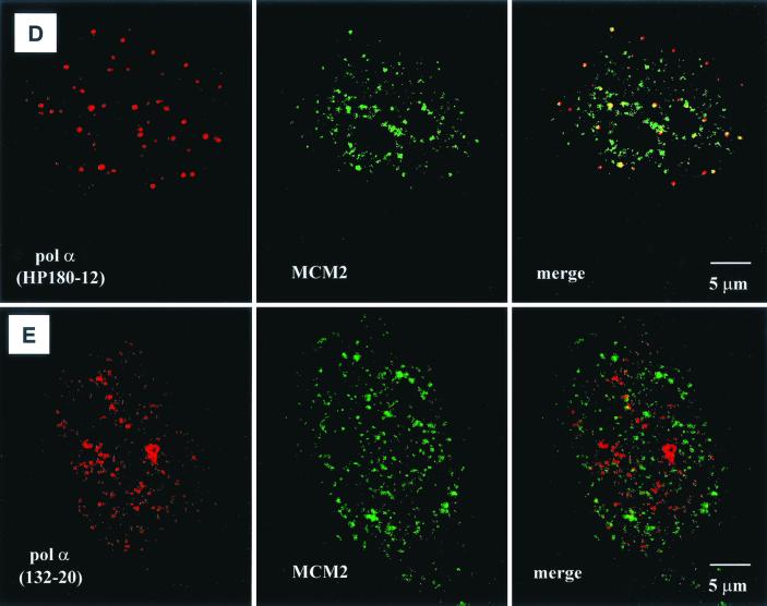
Localization of SJK132-20- and HP180-12-reactive Pol-Prim populations in replicating CV-1 cells by confocal microscopy. (A) Phosphorylated Pol-Prim colocalized with sites of active DNA synthesis in S phase cells identified by BrdU incorporation (right site, upper panel). Accumulations of the SJK132-20-reactive and therefore phosphorylated Pol-Prim (right site, lower panel) colocalized with the BrdU signal (left site). (B) Hypophosphorylated Pol-Prim detected with anti-p180 monoclonal antibody HP180-12 (right site, lower panel) does not colocalize with sites of active DNA synthesis in S phase cells (right site, upper panel). The merged image showed that hypophosphorylated Pol-Prim is not present at sites of DNA replication (left site). (C) Late-S-phase cells, identified by an increased number of large BrdU foci (right site, upper panel), showed a sharp reduction of the HP180-12 signal (right site, lower panel, and left site, merged image). (D) Hypophosphorylated Pol-Prim colocalized with MCM2 in early-S-phase cells. The merged image showed 50% colocalization of HP180-12-reactive Pol-Prim with the MCM2 speckles (right panel). (E) The merged image illustrates that the phosphorylated Pol-Prim (SJK132-20 reactive) does not colocalize with MCM2 (right panel).


