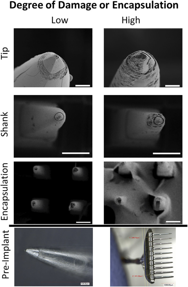
FIGURE 5.

Tip and shank damage occurred on some implanted electrodes and encapsulation occurred on four implanted arrays. Representative high magnification images of undamaged/unencapsulated and damaged/encapsulated electrodes. Tip images were taken from P1 array 1, with the degraded tip showing demetallation and biologic fouling (scale bar is 10 µm). Shank images were taken from P2 lateral stimulating array (scale bar is 100 µm). The degraded shank shows multiple surface and subsurface irregularities including pitting and delamination from the tip. Encapsulation images were from P2 medial stimulating array (scale bar is 100 µm). Pre implant optical images are provided to display pristine electrodes.
