Figure 1.
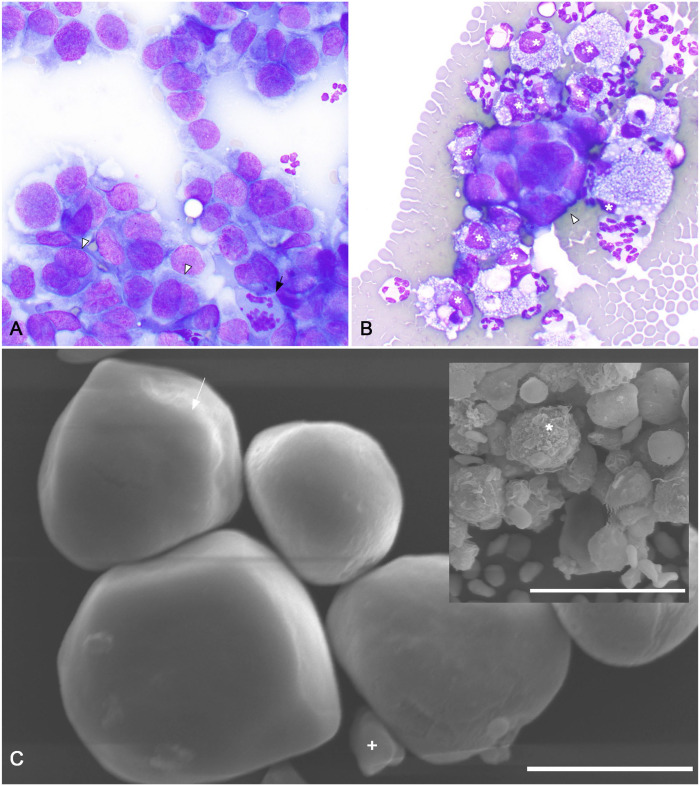
Antemortem fine-needle aspirate biopsy and peritoneal effusion cytocentrifuge and scanning electron microscopic findings of a feline sarcomatoid renal cell carcinoma (SRCC) with neoplastic peritoneal effusion. A. Cytologic features of a fine-needle aspirate biopsy of the SRCC. Large (~15–25-µm diameter), rounded neoplastic cells are arranged in loosely cohesive branching formations. Anisocytosis, anisokaryosis, and nuclear:cytoplasmic ratios are moderate. Cells borders are ruffled and indistinct. Nuclei are irregularly rounded to ovoid with finely stippled chromatin and, occasionally, a single large prominent nucleolus. A few bi- and trinucleate cells with frequent nuclear molding were observed (arrowheads). Within individual intact multinucleate cells, nuclei sometimes varied moderately in size. A few, sometimes aberrant, mitotic figures were present (arrow). Modified Wright stain. 500×. B. Cytocentrifuge preparation of peritoneal effusion. Occasional, variably sized, 3-dimensional clusters of neoplastic cells (arrowhead) are present. Cytomorphology of these cells is similar to that of the neoplastic cells from the fine-needle aspirate biopsy of the left perihilar SRCC. Heavily vacuolated macrophages (asterisks), nondegenerate neutrophils, and many erythrocytes are also present. Modified Wright stain. 1,000×. C. Scanning electron microscopy of cells within the neoplastic peritoneal effusion. Rare, small, loosely interlocking aggregates of polygonal-to-rounded cells 15–20-μm diameter are present. These cells have diffusely smooth cell surfaces, sometimes with a subtle leafy ridge (arrow). A centralized, cytoplasmic bulge compatible with a centrally located nucleus is present. A few smooth-surfaced, discoid, ~5-μm diameter erythrocytes were also observed (+). Bar = 10 µm. Inset: activated macrophages with irregular surfaces with multiple ridges, valleys, and indentations are also present (asterisk). Bar = 30 µm.

