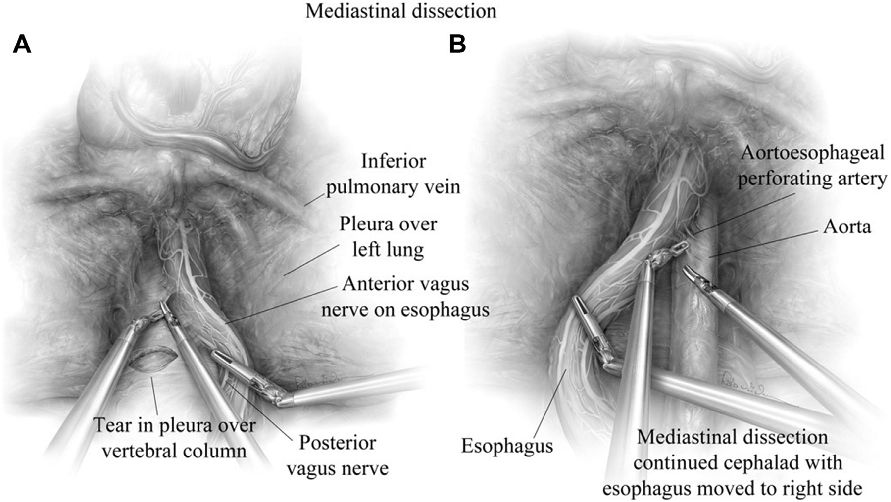
Fig. 5.

(A) Mediastinal dissection is continued until the inferior pulmonary vein is visualized. The left vagus nerve is readily identified and traced to the anterior vagus along the esophagus. (B) During anterior and posterior dissection, the mediastinal borders should always be well visualized to reduce injury to the aorta, vagus nerves, pleurae, vertebral column and the esophagus. (From Karush J, Sarkaria IS. Robotic-assisted giant paraesophageal hernia repair and Nissen fundoplication. Oper Tech Thorac Cardiovasc Surg. 2013;18(3):209; with permission.)
