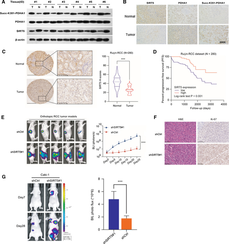
Fig. 6.
SIRT5 correlates with hyposuccinylation and progression of ccRCC. The levels of succinylated PDHA1 and SIRT5 were compared in 6 paired tumor and normal tissues using western blotting (A) and immunohistochemistry (B). C Representative images of immunohistochemical staining for SIRT5 protein in the Ruijin-ccRCC dataset. SIRT5 expression in tumor and normal tissues from the Ruijin-ccRCC dataset. D Kaplan–Meier analysis of PFS of patients stratified by SIRT5 expression in the Ruijin-ccRCC dataset. In vivo BLI (E) and HE staining (F) of an orthotopic model generated with WT and SIRT5-KD Luc-RENCA cells. G In vivo BLI of a lung metastasis model generated with WT and SIRT5-KD Luc-Caki-1 cells

