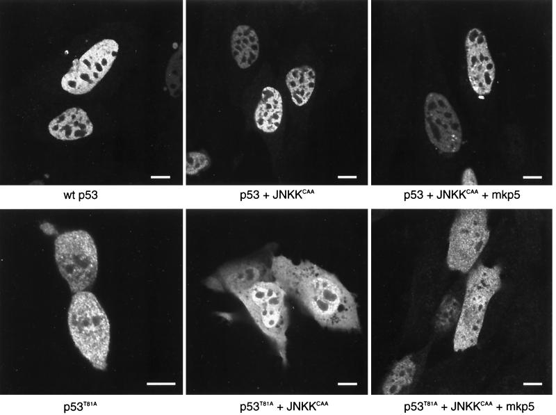
FIG. 6.
Cellular localization of wt and T81A p53 forms. p53 null 10.1 cells were transfected with wt p53, p53 plus JNKKCAA, p53 plus JNKKCAA plus MKP5, p53T81A, p53T81A plus JNKKCAA, or p53T81A plus JNKKCAA plus MKP5. Cells were immunolabeled with a rabbit polyclonal antibody to p53 (FL-393) (Santa Cruz Biotechnology) (diluted 1:500) followed by detection with fluorescein-conjugated anti-rabbit immunoglobulin G (heavy plus light chains) (Vector Laboratories). Cells were illuminated with the 488-nm line of the argon laser of a Leica TCS-SP (UV) confocal laser scanning microscope and examined using a 100×, 1.4 -numerical-aperture objective lens. For wt p53 (top three panels), labeling is restricted primarily to the nucleus (excluding the nucleoli) and nuclear bodies. Little or no cytoplasmic labeling is seen. For p53T81A (bottom three panels), labeling is divided between the nucleus and the cytoplasm. The lower left panel shows a pair of cells that are completing division. In the lower right panel, p53T81A is divided evenly between the nucleus and the cytoplasm in cells that express the protein at higher levels; at lower expression levels, it is found primarily in the nucleus. In all cases, p53 labeling is restricted from the nucleolus. Bars, 10 nm

