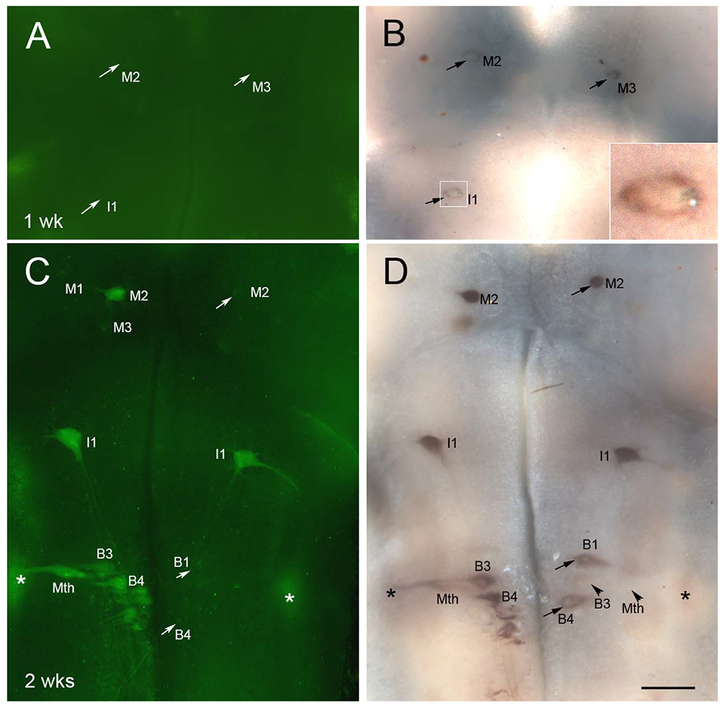
Figure 11.

Neurons in which caspases are activated post transection first upregulate PTPσ mRNA expression. A-D: Lamprey brains were stained for activated poly-caspases (A,C) followed by wholemount ISH for PTPσ mRNA (B,D), at 1 week (A,B) and 2 weeks (C,D) after SC transection. Neurons undergoing apoptosis were positive for activated caspases, as shown in C, and also expressed PTPσ mRNA, as shown in D. However, many neurons that were already slightly positive for PTPσ mRNA at 1 week post transection, e.g., M2, M3, and I1 in B, did not yet show caspase activation. Even at 2 weeks post transection, when PTPσ expression was strong, some PTPσ-expressing cells did not show caspase activation, e.g., M2, B1, and B4 in C and D (arrows), although most of these will eventually show caspase activation (Hu et al., 2013), indicating that expression of PTPσ mRNA precedes activation of caspases. In any one animal, some bad regenerators (e.g., B3, Mth, arrowheads in D) showed neither caspase activation nor PTPσ expression. Asterisks in C and D indicate the octavolateralis nucleus, containing the sensory neurons of the lateral line nerve. Insert in B is enlargement of I1. Scale bar = 250 μm in D (applies to A-D).
