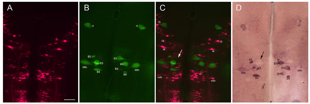
Figure 12.

PTPσ mRNA is expressed in neurons undergoing apoptosis but not in regenerating neurons. A: Regenerated spinal-projecting neurons retrogradely labeled with the fluorescent tracer DTMR applied to a second transection placed 5 mm caudal to the original lesion 7 weeks later. This identified those spinal-projecting neurons that had regenerated beyond the transection. B: Reticulospinal neurons (identities indicated) undergoing apoptosis are stained by FLICA in green. C: Overlay of images in A and B. The absence of double-labeled neurons indicates that neurons whose axons had regenerated do not show caspase activation and vice versa. D: ISH for PTPσ reveals that FLICA-positive neurons also expressed PTPσ mRNA. Variations in staining density may indicate different stages of apoptosis. An example is that a swollen B1 neuron was weakly stained with FLICA in C and negative for PTPσ in D. This could reflect that it was in an advanced stage of apoptosis (arrows). Scale bar = 100 μm in A (applies to A-D).
