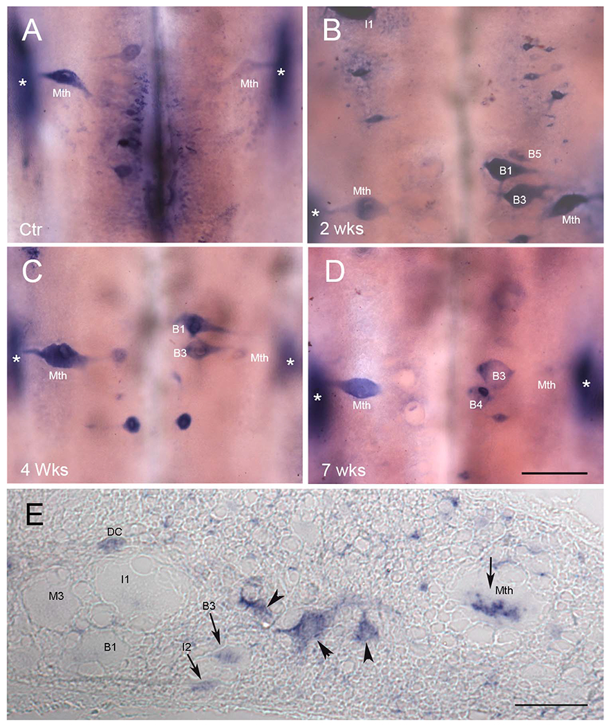
Figure 7.

PTPσ mRNA is expressed selectively in bad regenerators. ISH of PTPσ mRNA in whole-mounted lamprey brain shows expression mainly in neurons whose capacity to regenerate axons has previously been reported as weak. Expression was increased after SC transection, concomitantly with the increase in CSPG levels at the transection site. A: Control. B-D; B, 2 weeks, C, 4 weeks, and D, 7 weeks post transection. E: ISH of PTPσ mRNA in a transverse section of SC 2–3 mm rostral to the transection site at 2 weeks post transection. Some individual axons are labeled, based on the description of Rovainen et al. (1973). Arrowheads point to unidentified neurons. Arrows highlight axons that contain mRNA for PTPσ. DC, dorsal cell; Mth, Mauthner axon. In all frames, * indicates the locations of the octavolateralis nuclei, which are out of the focal plane. They expressed PTPσ, probably because they are subject to close axotomy during dissection of the brain at time of removal. See text. Scale bar = 200 μm in D (applies to A-D). Scale bar in E = 80 μm.
