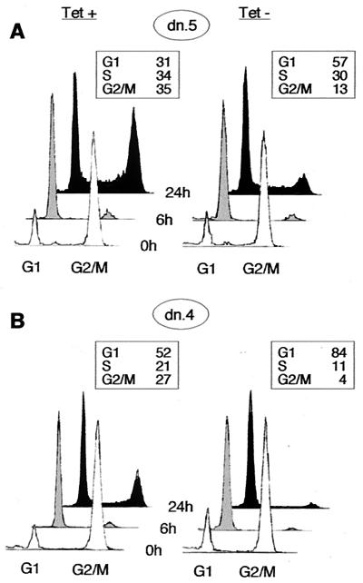
FIG. 3.
Inhibition of progression through G1 phase following induction of Cdk2-dn during a nocodazole block. Dn.5 (low expressor; A) and dn.4 (high expressor; B) cells were incubated with nocodazole for 24 h in the presence or absence of Tet. The mitotic cells were washed off the dish and replated in the absence of nocodazole (0 h; white profiles); cells were collected for flow cytometry at 6 h (grey profiles) and 24 h (black profiles) while maintaining the respective Tet conditions. Boxed areas show the percentage of cells in each fraction at 24 h.

