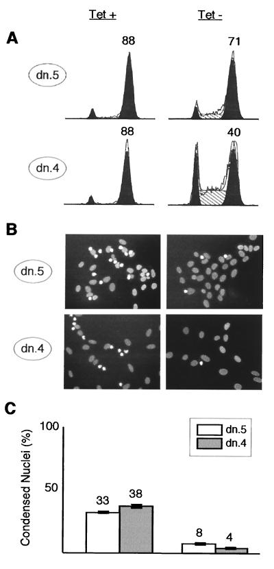
FIG. 8.
Cdk2-dn arrests cells in G2, prior to DNA condensation. Dn.5 and dn.4 cells were synchronized at the G1/S border by incubation with HU for 24 h in the presence (left) or absence (right) of Tet. HU was then removed, and nocodazole was added; 24 h after HU release, portions of each culture were either collected for flow cytometry or fixed and stained with bisbenzimide. (A) Flow cytometry profiles. G1 and G2/M fractions are outlined in black, S phase fractions are hatched, and the percentage of cells in G2/M is shown above that peak. (B) Representative fields with nuclear DNA stained by bisbenzimide. Interphase nuclei are broad, oval, and pale; early mitotic nuclei trapped by nocodazole are condensed, irregularly shaped, and bright. (C) Quantitation of the results in panel B, expressed as the percentage of nuclei in random high-power fields showing a condensed morphology. The bars depict mean numbers plus or minus ranges from two counts of more than 200 randomly chosen cells per condition. Similar results were obtained in a second experiment (not shown).

