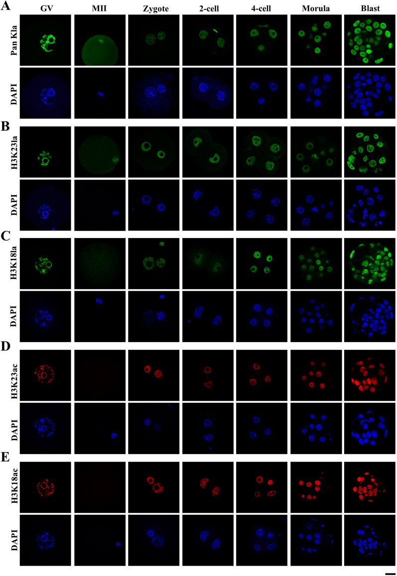
Fig. 1.
Nuclear accumulation of histone lactylation and histone acetylation in mouse oocytes and pre-implantation embryos. a–e Histone lactylation and histone acetylation were detected in mouse oocytes and pre-implantation embryos. Specific antibodies were used to detect the pan histone lactylation (a), H3K23la (b), H3K23ac (c), H3K18la (d), H3K18ac (e) in mouse oocytes at the germinal vesicle (GV) and metaphase II (MII) stages, as well as pre-implantation embryos at the zygote (zygote), 2-cell (2-cell), 4-cell (4-cell), morula (Morula) and blastocyst (Blast) stages. Pan histone lactylation, H3K23la and H3K23ac were shown in green. H3K18la and H3K18ac were shown in red. The DNA was stained with DAPI (Blue). More than 9 embryos were examined in each stage of each condition. Scale bars: 20 μm

