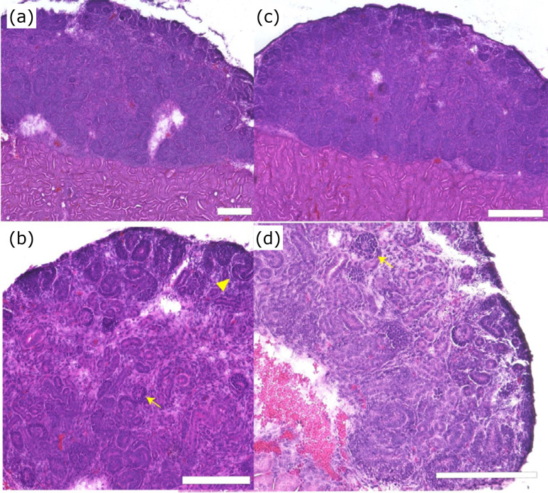
Figure 6. Effect of spinal cord on mouse metanephros reaggregation spheroid during transplantation. (a) HE stained image of mouse metanephros reaggregation spheroid transplanted alone (low magnification). (b) High magnification. (c) HE stained images of mouse spinal cord and mouse metanephros reaggregation spheroid transplanted alone low magnification. (d) High magnification. Scale bar: 200 μm. In the center of the tissue, glomeruli and multiple tubular structures can be observed. There were no significant differences between spheroids implanted with or without the spinal cord.
HE: hematoxylin and eosin; arrowhead: glomerulus; arrow: S-shaped body cap mesenchyme and S-shaped structures can be detected on the periphery of the spheroid.

