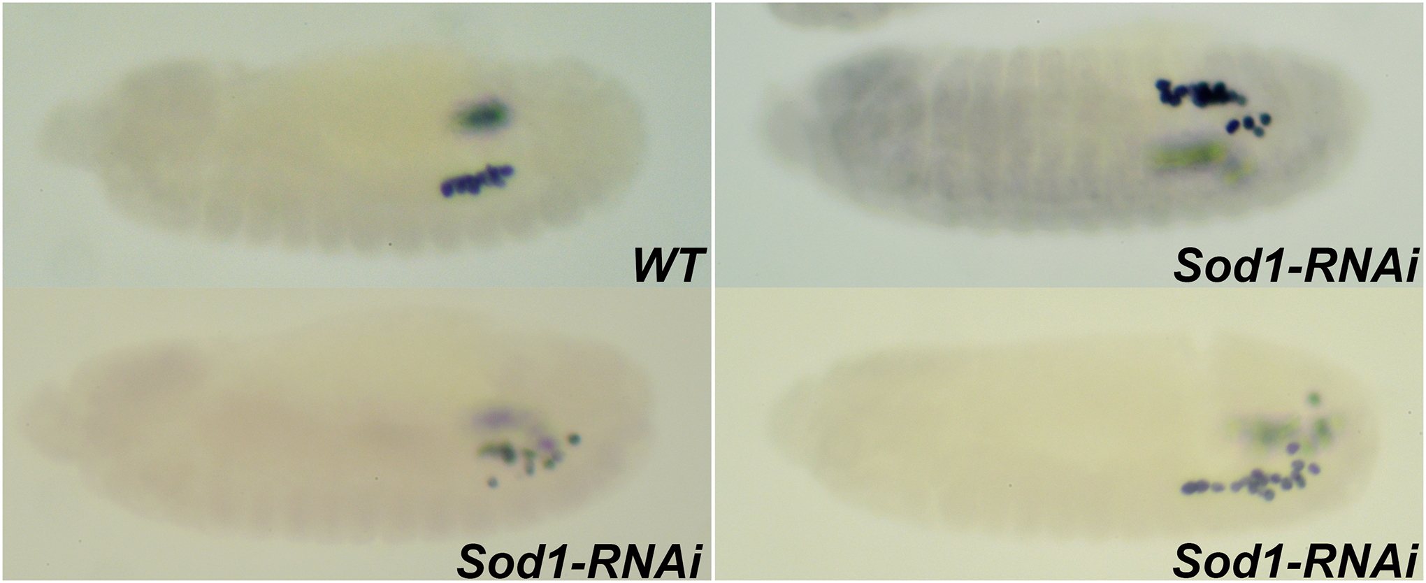
FIGURE 4. Embryos maternally compromised for Sod1 exhibit defects in PGC migration.

Stage 13 embryos compromised for Sod1 using RNAi show defective PGC migration as assessed by anti-Vasa labeling. Embryos are oriented anterior to the left and dorsal side on top in all the panels.
WT. At Stage 13, PGCs cluster and align against the SGPs situated in the para-segments 12 and 13.
Sod1-RNAi. MAT-Gal4/UAS-Sod1-RNAi embryos at stage 13 exhibit defects in PGC migration resulting in scattering of PGCs in the posterior of the embryo
