
FIGURE 5. PGCs from embryos compromised for Pxt display defects in PGC adhesion and migration at cellular blastoderm stage.
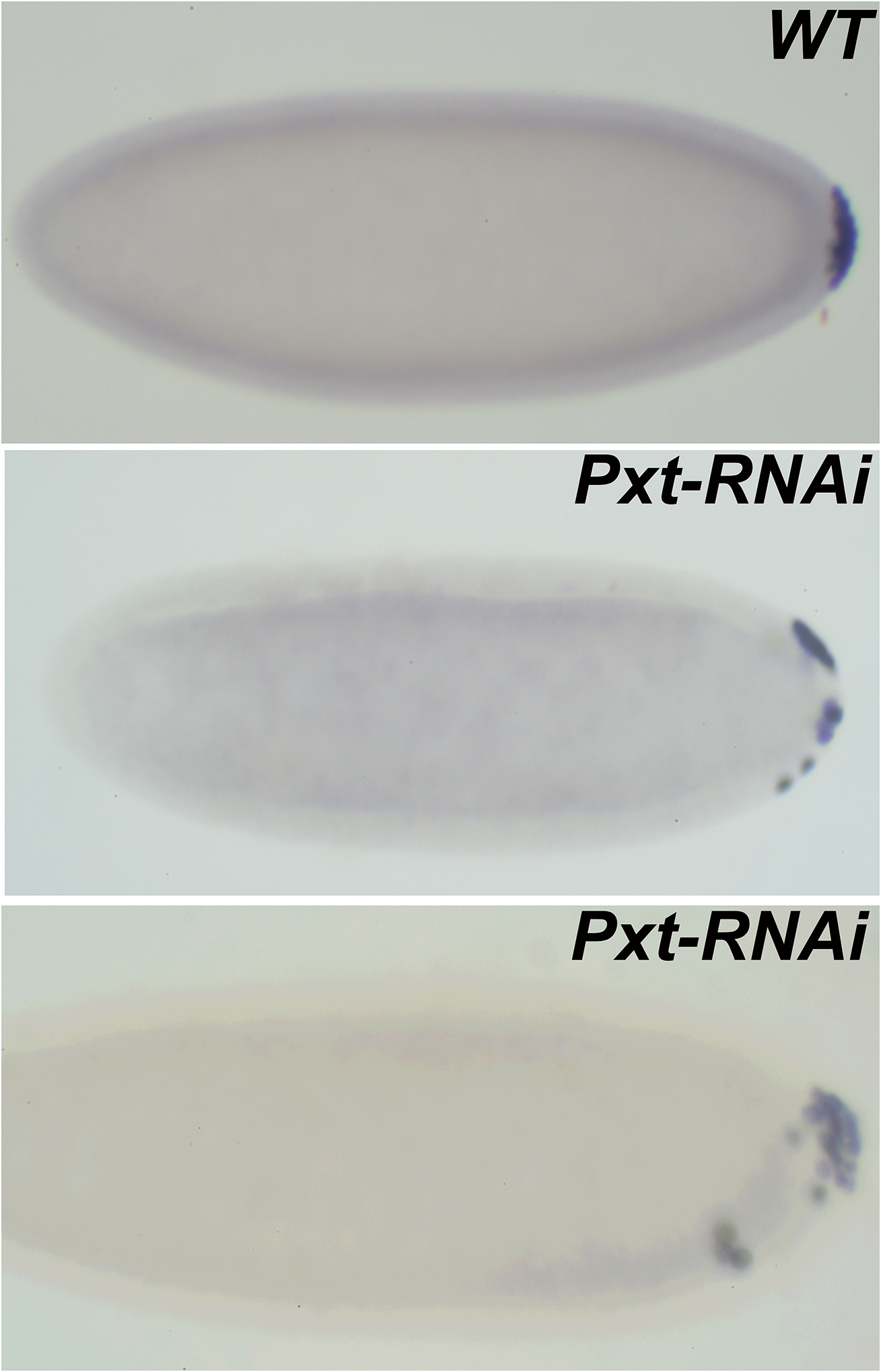
Embryos labeled with Vasa antibodies are oriented anterior to the left and dorsal side on top in all panels.
WT. PGCs in a cellular blastoderm stage embryo remain clustered at the posterior pole. Pxt-RNAi. In MAT-Gal4/UAS-Pxt-RNAi embryos pole cells lose adhesion and migrate away from the posterior pole. In more severe cases, PGCs not only migrate away from the pole but also invade the embryo. Interestingly, these pole cells move away from dorsal side and instead migrate on the ventral surface (bottom panel). Similar defects are also observed in Pxt mutant embryos collected from a viable, partial loss of function allele.