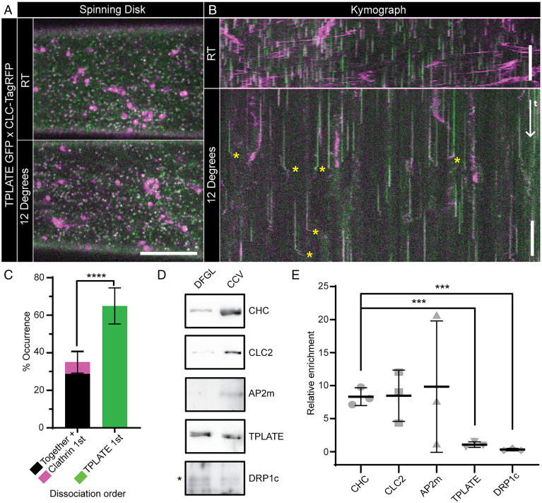
Fig. 1.
TPLATE is only loosely associated with CCVs. Example spinning-disk images (A) and kymographs (B) of Arabidopsis hypocotyl epidermal cells expressing TPLATE-GFP (green) and CLC2-TagRFP (magenta) at either room temperature (RT) or 12 °C. Yellow asterisks note example departure traces where CCVs are visible after dissociation from the PM. (C) Quantification of departure traces based on the order of departure (SI Appendix, Fig. S1B). n = 14 cells from independent plants, 258 departure traces. ****P < 0.0001, Student’s t test. Representative Western blots of endocytosis proteins during CCV purification (D) and quantification of proteins in the CCV fraction relative to an earlier purification step (DFGL) (E). n = 3 independent CCV purifications. ***P > 0.001, Student’s t tests compared to clathrin heavy chain (CHC). Plots, mean ± SD. (Scale bars, A, 5 µm; B, 60 s.)

