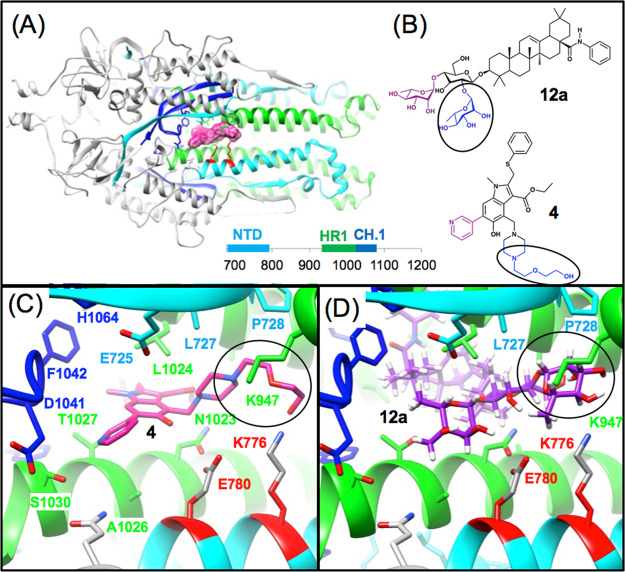
Figure 12.
Common structural features of predicted ARB derivative 4 and OA saponins bound to Site 1. (A) Ribbon model of the S2 segment, using color to highlight the NTD (cyan), HR1 (green), and CH.1 (blue) segments. (B) 2D structures of 4 and saponin 12a highlighting in blue and purple regions of overlap between the two 3D models. (C) Lowest energy binding mode of ARB derivative 4 shown in magenta highlighting residues E780 and K776 in red from the NTD segment which undergoes major pH-mediated conformational changes. (D) Predicted Structure of OA saponin 12a, where the common overlapping structural features of 4 and 12a are highlighted with a circle.

