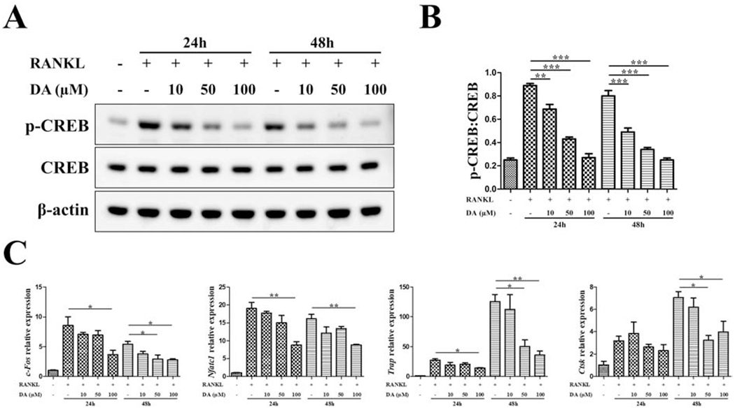
Fig. 2. CREB activity is inhibited by DA in osteoclastogenesis.

RAW cells were treated with indicated concentration of DA in the presence of 10 ng/mL RANKL. At different time points of osteoclastogenesis (24h, 48h), p-CREB and CREB level was detected by western blot (A) and quantitated as a ratio of p-CREB/CREB (B). (C) Relative expression of osteoclastic genes (c-Fos, Nfatc1, Trap, Ctsk) were determined by RT-qPCR; normalized to B2m. n=3 for all experiments; *P < 0.05, **P < 0.01, ***P < 0.001. Data shown as mean ± SEM.
