
Fig. 3. D2R-dependent inhibition on CREB activity in osteoclastogenesis.
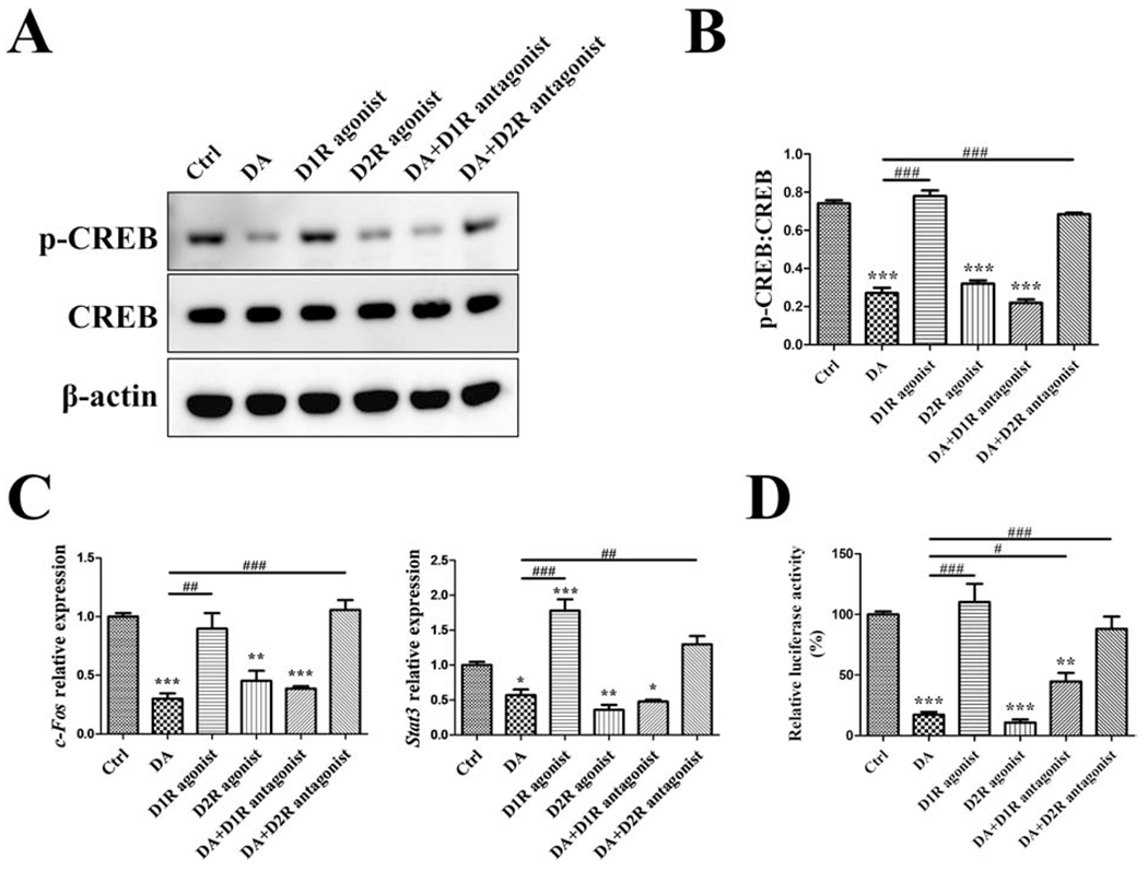
RAW cells were treated with vehicle (Ctrl), 100 μM DA, 100 nM D1R agonist SKF38393, 100 nM D2R agonist quinpirole, 100 μM DA plus 10nM D1R antagonist SCH23390 (after 30min SCH23390 pretreatment), or 100 μM DA plus l0nM D2R antagonist haloperidol (after 30min haloperidol pretreatment), in the presence of 10 ng/mL RANKL for 1 day. (A, B) Western blot detection of p-CREB/CREB. (C) RT-qPCR detection of c-Fos and Stat3 genes; normalized to B2m. (D) For p-CREB luciferase activity assay, p-CREB-Luc reporter-transfected RAW cells were used to receive the treatment same as above. n=3 for all experiments. *P < 0.05, **P < 0.01, ***P < 0.001 vs. Ctrl group; #P < 0.05, ##P < 0.01, ###P < 0.001 vs. DA group. Data shown as mean ± SEM.