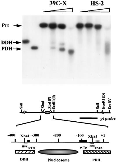
FIG. 3.
Configuration of DH sites in the heterochromatic transgene. Nuclei isolated from third instar larvae were incubated with DNase I at concentrations of 0, 0.03, 0.06, and 0.12 U/μl. Purified DNA was cleaved with SalI, and the fragments were analyzed by Southern blotting using the pt DNA fragment as the hybridization probe. Proximal (PDH) and distal (DDH) DH sites are indicated; the parental band, which is created by SalI digestion of the DNA not cleaved in nuclei by DNase I, is indicated (Prt). A partial restriction map of the hsp26 transgene is shown below: the (CT)n regions (black box), heat shock elements (HSE) (white box), the TATA box (gradient box), the PDH site (hatched box), the DDH site (hatched box), and the positioned nucleosome between the PDH and the DDH are diagrammed.

