. 2021 Dec 8;10:e72345. doi: 10.7554/eLife.72345

Figure 1. lratd2a-expressing neurons in the right dHb connect asymmetric pathway from the olfactory bulb to ventral IPN.

(A) Pattern of lratd2a expression at 5 days post fertilization (dpf), open arrowhead indicates right dHb and black arrowheads the bilateral vHb. (B) Sequences of WT (top) and transgenic fish (bottom) with QF2 integrated within the first exon of the lratd2a gene. PAM sequences are green, the sgRNA-binding site red and donor DNA blue. Confocal dorsal views of Tg(lratd2a:QF2), Tg(QUAS:mApple-CAAX) and Tg(lhx2a:gap-YFP) labeling in a (C-C’’) 5 dpf larva and in transverse sections of the adult brain at 3 months post-fertilization (mpf) at the level of the (D-D’’) dHb and (E-E’’) olfactory bulb. Axons of lhx2a olfactory mitral cells (open arrowheads, C and D) terminate at lratd2a dHb neurons. (F-F’’) Lateral view of Tg(lratd2a:QF2), Tg(QUAS:mApple-CAAX), Tg(gng8:GFP) larva at 6 dpf with mApple-labeled dHb terminals at the ventral interpeduncular nucleus (vIPN). Dorsal habenular nuclei (dHb), dorsal interpeduncular nucleus (dIPN). (G-G’’) Axonal endings of lratd2a dHb neurons are restricted to the ventralmost region of the vIPN in transverse section of 2.5 mpf adult brain. Scale bar, 50 μm. A-P, anterior to posterior; L-R, left-right; OB, olfactory bulb.

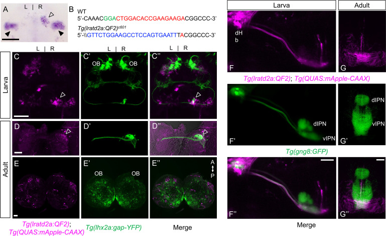
Figure 1.

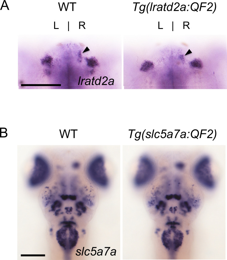
Figure 1—figure supplement 1. Targeted genomic integration does not disrupt endogenous gene expression.

Figure 1—figure supplement 1.

Expression patterns of (A) lratd2a and (B) slc5a7a are similar in 5 dpf wild-type and heterozygous transgenic larvae. Arrowheads point to the dHb. Dorsal views, scale bars are 100 μm.
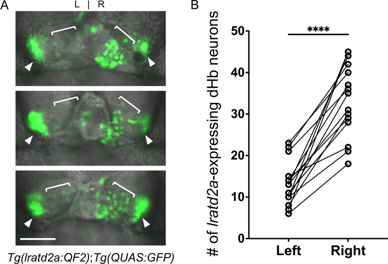
Figure 1—figure supplement 2. Asymmetry of lratd2a-expressing dHb neurons.

Figure 1—figure supplement 2.

(A) Dorsal views of the habenular region of three Tg(lratd2a:QF2); Tg(QUAS:GFP) larvae at 7 dpf. Each confocal image is a single focal plane (1 μm). Brackets indicate regions of lratd2a-expressing neurons in the dHb and arrowheads point to the bilateral vHb. Scale bar, 25 μm. (B) Quantification of lratd2a-expressing dHb neurons. Each line connecting the circles corresponds to a single larva. Mean is 13.1 ± 1.4 cells in the left dHb and 33.5 ± 2.3 cells in the right, n = 15 larvae [p < 0.0001, paired t-test].
Figure 1—figure supplement 2—source data 1. Source data associated with Figure 1—figure supplement 2B.
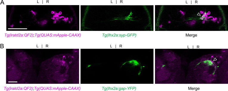
Figure 1—figure supplement 3. lhx2a-expressing olfactory cells innervate lratd2a dHb neurons.

Figure 1—figure supplement 3.

Axons of lhx2a olfactory mitral cells terminate at mApple labeled lratd2a dHb neurons. Dorsal confocal views of (A) Tg(lratd2a:QF2), Tg(QUAS:mApple-CAAX); Tg(lhx2a:syp-GFP) pre-synaptic labeling (open arrowhead) in a 5 dpf larva. The synaptophysin GFP-labeled pre-synaptic boutons are distributed over approximately 5% of the region occupied by lratd2a neurons. (B) Tg(lratd2a:QF2), Tg(QUAS:mApple-CAAX); Tg(lhx2a:gap-YFP) axon terminal labeling (open arrowhead) in a transverse section of the adult brain at three mpf. Scale bar, 50 μm.