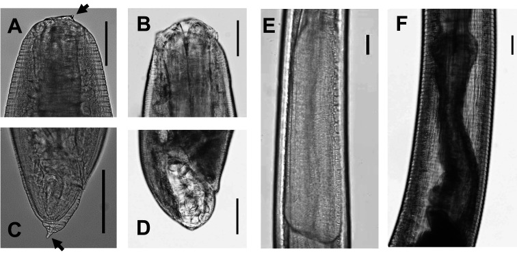
Fig. 3.
Morphology of third-stage (L3) and fourth-stage (L4) larvae of Anisakis simplex sensu stricto
A, Cephalic end of L3 larva (arrowed line: boring tooth); B, Cephalic end of L4 larva; C, Caudal end of L3 larva (arrowed line: mucron); D, Caudal end of L4 larva; E, Ventricular part of L3; F, Ventricular part of L4. Bar: 100 μm.

