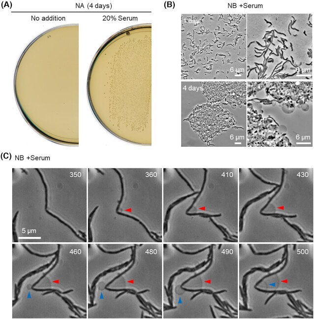
Figure 1.
Growth and morphology of S. moniliformis. (A) Growth of S. moniliformis type strain 9901 on NA plate containing 20% calf serum at 30°C. (B) Phase-contrast micrographs of S. moniliformis in NB with serum after 1 or 4 days of incubation at 30°C. (C)Streptobacillus moniliformis cells were cultured in NB with serum in the CellASIC ONIX microfluidic system for time-lapse microscopy. Individual frames are extracted from Video S1 (Supporting Information). Numbers in the top right corner of each frame represent time (min) elapsed in the video. Arrowheads represent spontaneous formation of L-form-like structures.

