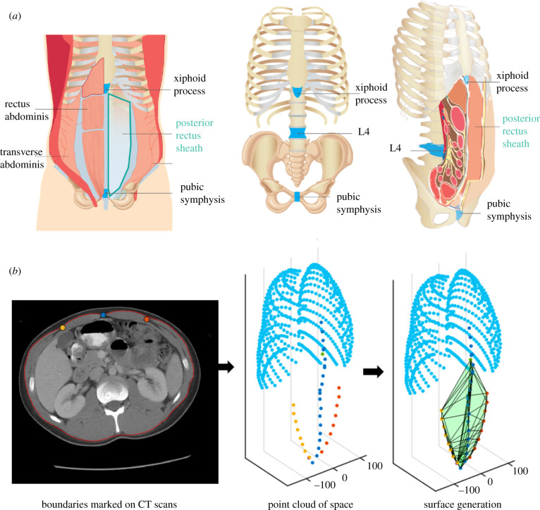
Figure 1.
Graphical representation of the PRSP and analysis used in this study. (a) Schematic of anatomy of the location of the PRSP enclosed by the xyphoid process (cranially), tip of the pubic symphysis (caudally), linea alba (medially) and semilunar line points (laterally). (b) Arbitrary participant showing the points from the segmentation from CT scans and the resulting computed space.

