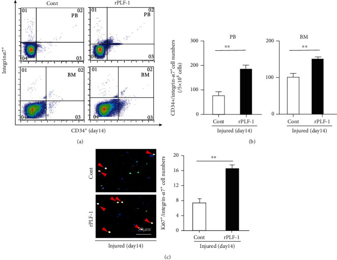
Figure 5.

rPLF-1 stimulated bone marrow (BM) MuSC production and mobilization in response to CTX injury. (a, b) Representative dot plots and quantitative data for the numbers of CD34+/integrin-α7+ MuSCs in BM and peripheral blood (PB). (c) After the isolation of BM-derived integrin-α7+ stem cells with magnetic beads, the cells were cultured on cover glasses for 24 hr and then subjected to double immunofluorescence with goat pAb against integrin-α7 (red) and mouse mAb against Ki67 (green). Representative double fluorescence images and quantitative data show the numbers of proliferating cells (×200 magnification). Results are mean ± SE (n = 7–8). ∗∗p < 0.01 vs. corresponding control groups by Student's t-test.
