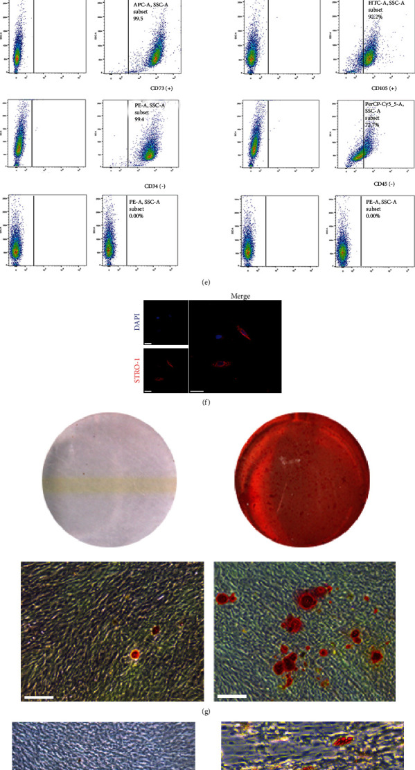
Figure 1.

Identification of PDLSCs. (a) Imaging examination of immature permanent teeth. (b) Scraping the middle third of the root of the tooth. (c) The morphology of primary PDLSCs (scale bar = 100 μm). (d) Morphology of the third-generation PDLSCs (scale bar = 100 μm). (e) Flow cytometry assay showed that PDLSCs were positive for CD29, CD73, CD90, and CD105, but negative for CD34 and CD45. (f) Immunofluorescence assay revealed that PDLSCs were positive for STRO-1 (scale bar = 50 μm). (g, h) PDLSCs had the potential to differentiate into osteoblasts and adipocytes (scale bar = 100 μm). On the left is the control group which was induced by complete medium. (i) PDLSCs had the potential to differentiate into chondrocytes (scale bar = 200 μm).
