FIGURE 1.

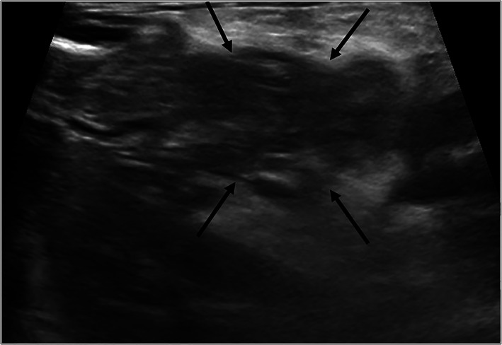
Ultrasonographic findings. Transverse plane B mode ultrasound image of the midbody of the pancreas (arrows). Note the hypoechoic pancreatic parenchyma with hyperechoic surrounding mesentery

Ultrasonographic findings. Transverse plane B mode ultrasound image of the midbody of the pancreas (arrows). Note the hypoechoic pancreatic parenchyma with hyperechoic surrounding mesentery