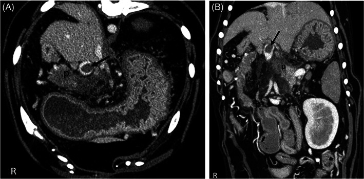
FIGURE 2.

Transverse (A) and dorsal (B) CTA image. (A) CTA venous phase transverse plane image of the cranial abdomen. Note the heterogeneously contrast enhancing pancreas (p) and oval thrombus in the portal vein (arrow). There is a large amount of fat stranding within the mesenteric fat surrounding the pancreas, indicating edema and inflammation. (B) CTA venous phase dorsal plane image of the cranial abdomen. Note the heterogeneously contrast enhancing pancreas (p) and oval thrombus in the portal vein (arrow)
