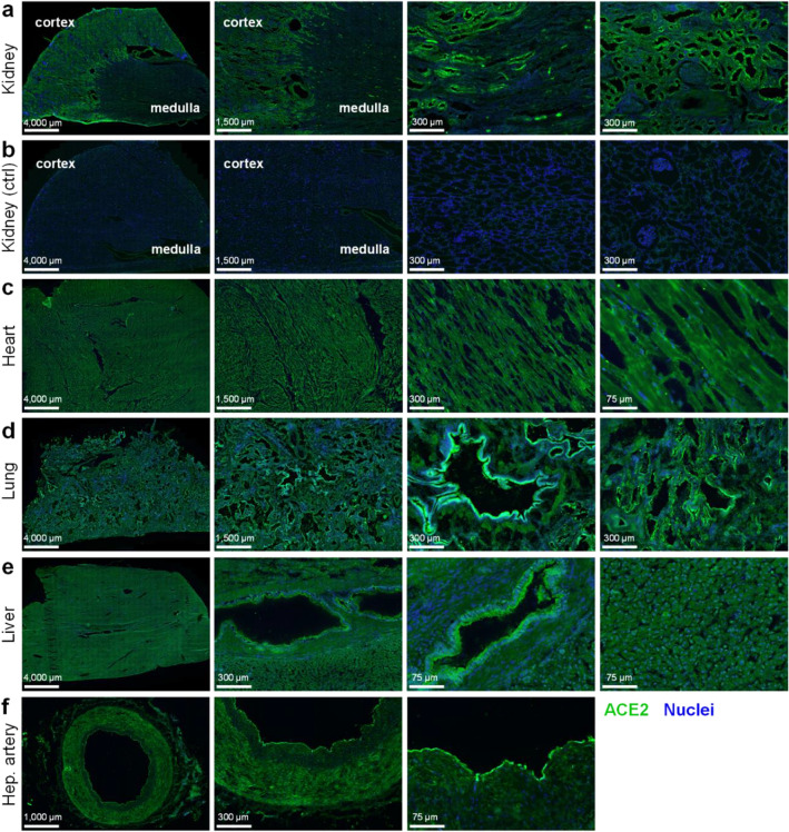
Figure 1.
Immunohistochemistry confirms positive staining for ACE2 in several human tissue types. Representative fluorescent images of 4% formaldehyde fixed human tissue sections (n ≥ 3 independent donors, stained in duplicate) treated with ACE2poly antibody (R&D AF933), visualised in green, and Hoechst 33342 nuclear stain, visualised in blue. Scale bars are as indicated in figure. Tissues shown include: (a) Kidney, with cortex and medulla indicated; (b) Kidney control section treated with secondary antibody and Hoechst 33342 alone; (c) Heart tissue, comprised predominantly of cardiomyocytes; (d) Lung, with preserved airway structures; (e) Liver, comprised predominantly of hepatocytes with preserved bile duct structures; (f) Hepatic artery section.

