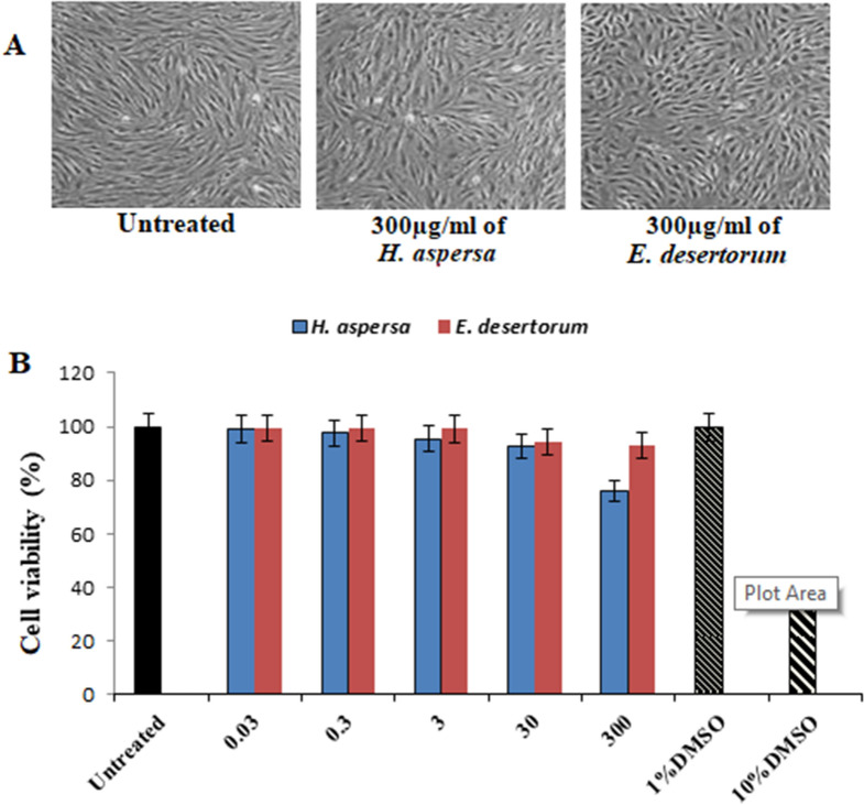
Figure 3.
Cytotoxicity evaluation of MEs of both selected snails. HSF cells were exposed to 300 µg/ml of MEs of both selected snails and cell viability was examined by SRB assay. (A) Representative images with magnification of (10 ×) taken by light microscopy of HSF cells untreated and treated with 300 µg/ml of both selected strains at 48 h. (B) Cell viability was calculated at 24, 48 and 72 h compared to untreated cells (control), DMSO (10%) and (1%) were used as positive and vehicle controls of cell death, respectively. Results represent the average of three independent experiments ± SD. *p < 0.05.

