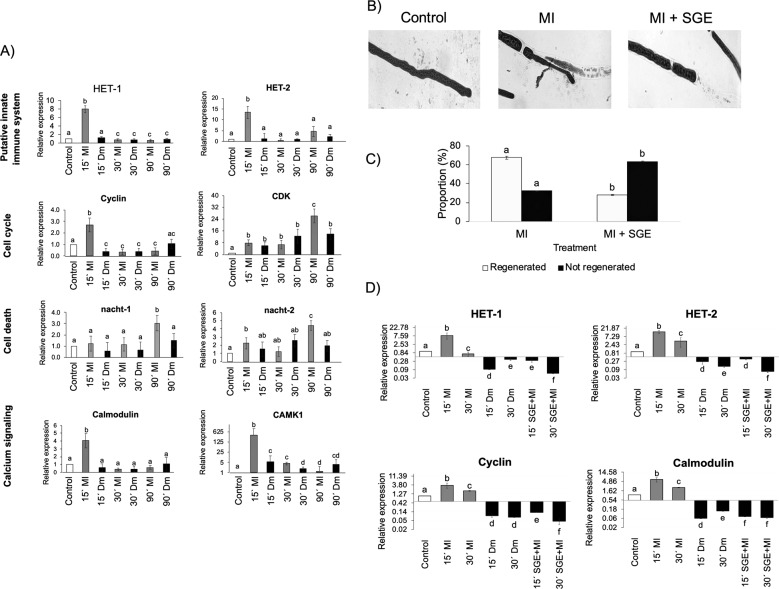
Fig. 4. Larvae salivary gland extracts block hyphal regeneration in T. atroviride.
A RT-qPCR gene expression analyzed of selected genes belonging to different processes related to hyphal regeneration. Bars indicate the relative expression level of the indicated gene in the control condition (white), after MI (gray), and after attack by D. melanogaster larvae (black). A–D Gene expression relative to the not challenged control (arbitrarily set to one), was calculated by the 2−ΔΔCT method. Different letters in the error bars indicate significant statistical differences with the control condition and constitutive control gene after ANOVA and Tukey tests (P > 0.05). Control: not challenged, undamaged T. atroviride. MI: After a mechanical injury. Dm: After D. melanogaster grazing. B Micro-photographs of a T. atroviride hypha (control), and injured with a clean scalpel MI, or a scalpel dipped in Salivary Gland Extract (MI + SGE). Black arrows in micro-photographs indicate the damaged hypha—scale bars (20 µm). C Hyphal regeneration after the indicated treatments represented as a percentage. Grey bars indicate the proportion of “Regenerated hyphae” and black bars the proportion of “Not regenerated hyphae”. Different letters above the bars indicate significant statistical differences between treatments (P < 0.05). Control: Control condition. MI: After a mechanical injury. D RT-qPCR of the genes het1, het2, cyclin, and calmodulin after MI + SGE treatment of T. atroviride hyphae or grazing by D. melanogaster larvae (black bars), mechanical injury (MI, gray bars), and control condition (white bars). Control: not challenged, undamaged T. atroviride. Dm: After D. melanogaster grazing. SGE + MI: After mechanical injury with a scalpel soaked in salivary gland extract. MI: Mechanical injury.

