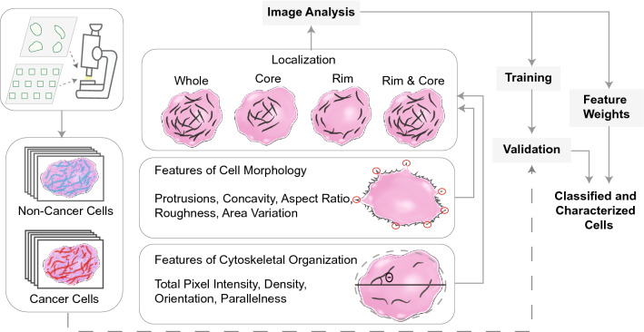
Figure 1.
An automated image processing framework quantifies features of cellular cytoskeletal and morphological structure from single cell images. These features were used to train parameters of a classification model and its performance was evaluated using validation data. The algorithm was able to accurately discriminate cancer cells from non-cancer cells and identify individual features that had the greatest influence on classification outcome.

