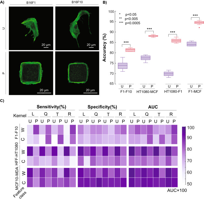
Figure 4.
Example of microcontact printing-based image analysis. (A) Comparison of B16-F1 and B16-F10 murine melanoma cells when unpatterned (U) or patterned (P) in 900 μm2 islands. (B) Improvement of pairwise predictive accuracy when hard-to-discriminate cell lines are patterned. (C) Sensitivity, Specificity, and AUC of physiologically relevant comparisons B16-F1/B16-F10, HFF-1/HT-1080, and MCF10A/MDA-MB-231 for both patterned (P) and unpatterned (U) cells across both whole (W) and core (C) classes and all four SVM kernels.

