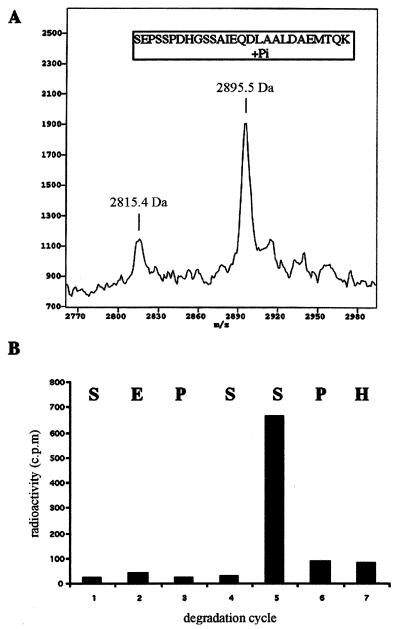
FIG. 6.
Determination of the precise ERK phosphorylation site of PLC β1. (A) Mass spectra of the peptides for HPLC fraction 36. The peptide samples were mixed with α-cyano-4-hydroxycinnamic acid. The mixture was applied to mass analysis using a G2025A MALDI-TOF mass spectrometer as previously described (51). (B) Phosphate release analysis of the 32P-labeled peptide. An aliquot of HPLC fraction 36 (∼1,000 cpm) was subjected to N-terminal sequencing, and 32P released from each cycle was analyzed for radioactivity. The corresponding amino acid residues released from each cycle are shown at the top of the chart.

