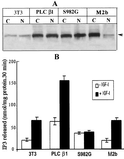
FIG. 7.
The responsiveness of wild-type PLC β1 and PLC S982G mutant or M2b to IGF-I stimulation in Swiss 3T3 cells. (A) Western blot analysis of nuclear PLC β1 from wild-type 3T3 cells and 3T3 cells overexpressing wild-type PLC β1, the PLC β1 S982G mutant, or the mutant M2b. The stable transfectants were selected as described in the text. Sixty micrograms of cytoplasmic proteins or 20 μg of nuclear proteins from these cells was separated by SDS–8% PAGE and probed with anti-PLC β1 MAb. (B) Nuclei from cells were analyzed for PLC activity as described in the text. The results are presented as the means ± standard deviations (n = 4). Note that the figure shows the result of typical experiments, and similar results were obtained from at least another two independent transfectants that express wild-type PLC β1, the S982G mutant, or M2b.

