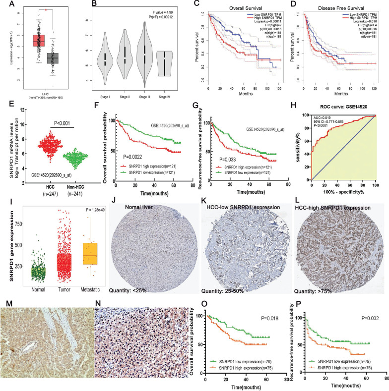
Figure 2.
High mRNA and protein expression of SNRPD1 correlates with poor prognosis in HCC patients. (A) The SNRPD1 expression level in HCC samples was significantly higher than normal liver tissues in the GEPIA database(P<0.05). (B) The SNRPD1 expression in HCC samples was incrementally upregulated with increasing tumor stages in the GEPIA database. (C-D) High SNRPD1 mRNA expression correlates with poor overall survival (C) and diseases free survival (D) of HCC patients in the GEPIA database. (E) SNRPD1 expression in HCC tissues was significantly higher than adjacent normal liver tissues in the GSE14520 dataset. (F-G) High SNRPD1 mRNA expression correlates with poor overall survival (F) and recurrence-free survival (G) of HCC patients in the GSE14520 dataset. (H) The receiver operating characteristic (ROC) curve revealed that SNRPD1 expression has a significant diagnosis value on HCC (AUC=0.819, P<0.001). (I) The SNRPD1 mRNA expression was incrementally upregulated in the normal, tumor, and metastatic tissues of HCC patients in the TNMplot database (https://www.tnmplot.com/). (J-L) Representative images of immunohistochemical (IHC) staining of SNRPD1 protein expression in normal liver tissues (J, expression quantity <25%), low expression HCC tissues (K, expression quantity 25-50%), and high expression HCC tissues in the Human Protein Atlas database (L, expression quantity >75%). (M-N) Representative image of IHC staining of SNRPD1 low (M)/high (N) protein expression in tumor tissue from 154 patients with HCC (x200 magnification). (O-P) High SNRPD1 protein expression correlates with poor overall survival (O) and diseases free survival (P) of 154 HCC patients.

