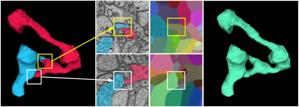
Fig. 2.
Mean embedding agglomeration (Sec. III-C). Shown here is a real example of self-contacting axon from the validation set. Left: a false split error (yellow box) in the Mutex Watershed [17] segmentation caused by a self-contact (white box). Middle top: PCA visualization reveals homogeneous embeddings across the false split. Middle bottom: each side of the self-contact receives distinct embedding vectors (white box). Note that the both sides appear to be distinct objects in the limited local context. Right: mean embedding agglomeration correctly heals the false split.

