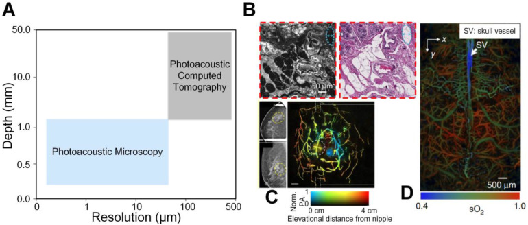
Figure 1.
(A) A continuum from micro to macro by photoacoustic imaging. (B) An example of micro-scale structural photoacoustic imaging. Photoacoustic microscopy and hematoxylin and eosin-stained figures of normal human breast tissue 17. (C) An example of macro-scale structural photoacoustic imaging. Photoacoustic computed tomography of human breast cancer 18. (D) An example of micro-scale functional photoacoustic imaging. Oxygen saturation of mouse brain 19. Adapted with permission from 17-19. Copyright © 2017, Copyright © 2018, and Copyright © 2015 Springer Nature.

