
Figure 1.
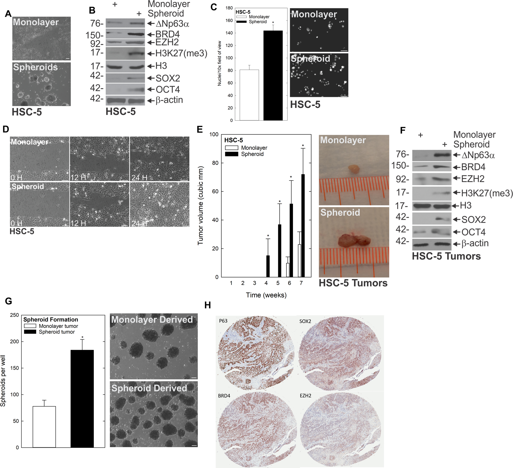
A subpopulation of SCC cells display traits of CSCs. A HSC-5 monolayer cultures maintained in growth medium were harvested and plated at 4×10⁴ cells per 10 cm dish in monolayer cultures or in non-attached conditions as spheroids and monitored for 10 d. Bars = 200 μm B At 10 d, lysates were electrophoresed for detection of the indicated epitopes. C At 10 d, monolayer and spheroid cells were trypsinized and single-cell suspensions were seeded onto Matrigel-coated membranes in Millicell chambers for invasion assays, or D replated as monolayer cultures and allowed to reach confluence at which time they were scratched with a 10 μl pipette tip to create a wound; wound closure was monitored over time. E HSC-5 spheroid- or monolayer-derived cells were injected subcutaneously at 1×10⁶ cells per site in nude mice and tumor growth was monitored. Tumor volume was determined by caliper measurements. The values are mean ± SEM (n=13). Asterisks indicate significant differences in tumor size between the spheroid and monolayer groups at each time point. F Protein extracts were prepared from tumors for immunoblotting to detect the indicated epitopes. G Monolayer- and spheroid-generated tumors were dissociated to create single-cell suspensions, and tumor cells were seeded for spheroid growth assays; spheroid number was monitored for 9 d. H Patient-derived SCC tumor arrays were immunostained to detect the indicated epitopes, and quantified.