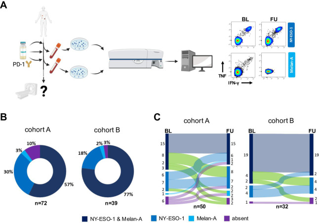
Figure 1.
Determination of functional TAA-reactive T cell profiles in the peripheral blood of patients with stage IV melanoma under PD-1 blockade. (A) Study design and experimental workflow. (B) Distribution of NY-ESO-1-reactive and Melan-A-reactive (dark blue), NY-ESO-1-reactive (blue) or Melan-A-reactive (light blue) T cell populations or the absence (purple) of both populations before the start of PD-1 blockade in both cohorts. (C) Proportions of circulating NY-ESO-1-reactive/Melan-A-reactive T cell populations under PD-1 blockade in either cohort. Green highlight indicates patients with a loss of at least one TAA-reactive T cell population under therapy. BL, baseline; FU, follow-up; IFN-γ, interferon gamma; PD-1, programmed cell death protein 1; TAA, tumor-associated antigen.

