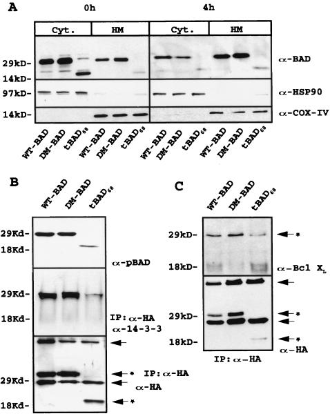
FIG. 4.
(A) BAD (WT, mutant, and truncated) expression in subcellular fractions of retrovirus-infected parental 32Dc13 cells. Western blots show BAD levels in mitochondrion (HM)- and cytoplasm (Cyt)-enriched fractions from parental cells in the presence of IL-3 or 4 h after its removal (upper gels). Anti-BAD (C-20) antibody (α-BAD) was used as the probe. Levels of the cytoplasmic marker HSP90 (middle gels) and mitochondrial marker subunit IV of cytochrome oxidase (COX-IV, lower gels) were measured as a control for equal loadings and the purity of subcellular fractions. Results are representative of three separate experiments. (B) Serine phosphorylation and 14-3-3 interaction of WT, DM56/61, and tBAD68 HA-BAD in retrovirus-infected parental cells. Western blot analyses were carried out with a mix of anti-pSer112 and -pSer136 antibodies to monitor phosphorylated BAD (upper gel). To study the interaction between 14-3-3 protein and WT, DM56/61, and tBAD68 HA-BAD, cell extracts were immunoprecipitated with the anti-HA antibody and blotted with anti-14-3-3 antibody (middle gel). Levels of WT, DM56/61, and tBAD68 HA-BAD were measured as a control for equal loadings (lower gel). Results are representative of three separate experiments. Arrows indicate heavy- and light-chain immunoglobulins; arrows with asterisks indicate antibody-specific reactive bands. IP, immunoprecipitate. (C) Interaction of HA-BAD (WT, mutant, and truncated) with BCL-XL in retrovirus-infected 32Dc13 cells. Anti-HA immunoprecipitates from WT, DM56/61, and tBAD68 HA-BAD retrovirus-infected 32Dc13 cells were blotted with anti-BCL-XL antibody (upper gel). Levels of HA-tagged protein were measured as a control for immunoprecipitation (lower gel). Results are representative of three separate experiments. Arrows indicate heavy- and light-chain immunoglobulins; arrows with asterisks indicate antibody-specific reactive bands.

