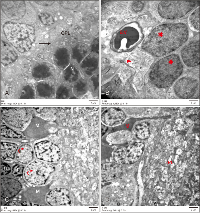
Fig. 6.
Electron micrograph of (T1DM+ insulin+hesp) group, scale bar=2 µm (A: ×1,500; B: ×2,000; C, D: ×1,200). (A) Showing preserved ultrastructure of most layers of retina. Rods with electron dense nuclei (N) and outer plexiform layer (OPL) with normal thickness an organization (arrow). (B–D) Inner nuclear layer containing many neurons with electron lucent cytoplasm (arrowheads) and euchromatic nuclei (curved arrows). Other neurons appeared with heterochromatic nuclei (star). Microglia (g), retinal blood vessel with normal basement membrane (B.V), Muller cells and inner plexiform layer (IPL) are seen. T1DM, type 1 diabetes mellitus; hesp, hesperetin 7-rhamnoglucoside.

