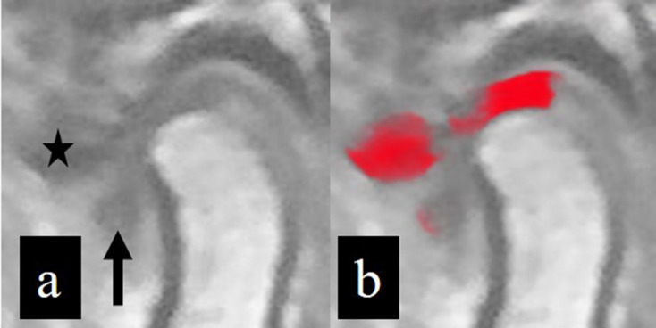
Figure 5.

Inputted (a) and outputted (b) image data. The disc situated superior to the condylar head is well segmented, but the anterior disc displacement area (arrow) cannot be identified; this indicates a false-negative result. The cortical area of the articular eminence (star) is erroneously identified as a disc area, indicating a false-positive result.
