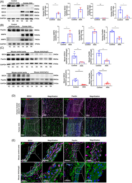
FIGURE 1.

SDC4 was reduced in human and mouse abdominal aortic aneurysm (AAA) tissues. (A and B) Representative western blots of SDC1–4 (A), paxillin, MMP2 and MMP9 (B) and densitometric analysis of human AAA and adjacent control tissues (n = 3). (C) Representative western blots of SDC4 and paxillin and densitometric analysis of abdominal aortas from AngII‐induced AAA mice, CaCl2‐induced AAA mice and control mice (n = 5). (D) Representative immunofluorescence images of SDC4 and paxillin in human AAA and adjacent control tissues (n = 3), α‐SMA (green), SDC4 and paxillin (red), and DAPI (blue) (scale bars, 250 μm and 100 μm) (magnified photographs). (E) Representative immunofluorescence images of SDC4 and paxillin in control and AngII‐induced AAA mouse samples (n = 5); α‐SMA (green), SDC4 and paxillin (red), and DAPI (blue) (scale bars, 300 μm and 100 μm) (magnified photographs)
